A Guide to Medical Imaging: CT, MRI, PET, PET-CT—Which One to Choose?


CT, MRI, PET, and PET-CT are common types of medical imaging used to detect and diagnose abnormalities in the body's internal structures or functions. However, what sets each imaging method apart? How do they differ in terms of principles and application? Here, we provide a comprehensive breakdown of what CT, MRI, PET, and PET-CT are all about.
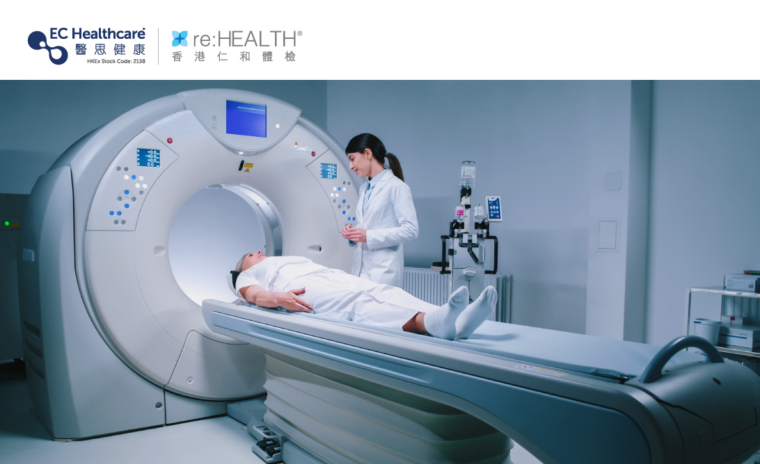
CT, Computed Tomography
CT uses X-ray radiation to capture multiple cross-sectional images of the body from different angles. X-rays pass through tissues with varying densities, creating detailed images of these cross-sections. Advanced computer technology reconstructs these images into clear and visible 3D images.
CT scans are commonly used for imaging the brain, heart, chest, abdomen, pelvis, and limbs. Doctors utilize the 3D images obtained from the scans to diagnose and monitor conditions such as tumors, fractures, osteoporosis, arthritis, brain injuries like strokes and hemorrhages, cardiac abnormalities, pneumonia, emphysema, abdominal and pelvic diseases including internal bleeding and intestinal obstruction. However, CT scans have lower sensitivity for soft tissues. Therefore, when soft tissue evaluation is required, patients may need to receive a contrast agent injection prior to the scan to enhance the clarity of the imaging results.
MRI, Magnetic Resonance Imaging
MRI is a radiation-free medical imaging method. It utilizes a strong magnetic field to temporarily align and rearrange hydrogen atoms in the body. The nuclei of these atoms emit energy in the form of radio waves, which are then detected by a computer. Through computational analysis, information regarding tissue structures, blood flow, and water distribution can be obtained. MRI is commonly used to examine the brain, heart, spine, intervertebral discs, nerve roots, spinal cord, muscles, bones, joints, and various abdominal organs and blood vessels, providing insights into their function and structure.
MRI, Magnetic Resonance Imaging, provides clear visualization of both hard and soft tissues, making it valuable in detecting tissue abnormalities. It plays a significant role in diagnosing benign or malignant tumors, determining cancer stages, monitoring treatments, identifying fatty liver, and assessing cartilage changes. However, MRI has limitations in detecting certain diseases such as blood cancer, esophageal cancer, gastric cancer, colorectal cancer, lung diseases, liver conditions, adrenal gland disorders, and prostate issues. Additionally, MRI is not suitable for pregnant women or individuals with implanted metal devices such as pacemakers, stents, or artificial heart valves.
PET, Positron Emission Tomography
PET is currently the most advanced and effective method for evaluating tumors. By injecting a radioactive isotope medication, which distributes in specific organs or tissues within the body, PET detectors can track and collect the dual positron energy generated by the medication, constructing a 3D image. PET is typically used for individuals suspected or diagnosed with head and neck cancer, esophageal cancer, lymphoma, breast cancer, lung cancer, colorectal cancer, and rectal cancer. It helps identify abnormally active tissues, track early cancer cell changes, differentiate tumor types, sizes, metabolic activity, and detect treatment effectiveness.
Additionally, PET is also useful for detecting and assessing diseases related to brain function, brain neurotransmission, myocardial blood flow, and cardiac function. This includes conditions such as schizophrenia, depression, Alzheimer's disease, cerebrovascular disorders, coronary artery disease, and myocardial infarction.
PET-CT, Positron Emission Tomography-Computed Tomography
PET-CT combines the advantages of PET and CT imaging techniques. By performing PET and CT scans simultaneously, it generates high-resolution images that combine cellular metabolism and tissue structure. Through PET-CT imaging, doctors can accurately identify early cancerous changes, determine the location and activity of tumors, and assess signs of spreading. It also aids in guiding biopsies, identifying radiation therapy targets, and detecting signs of recurrence, with an accuracy rate of over 90%.
Each of the aforementioned medical imaging techniques has its own advantages and limitations. Doctors will determine the appropriate and suitable type of medical imaging based on the individual's specific health condition. For more information, please consult your doctor.
Related Brands